Microscopy Particle Size Analysis
Characterize particle count, size, and shape
Why the big fuss over these little particles?
Particulate contamination in injectable drug products can cause undesired immune responses, vascular blockages, and a range of other complications. Because of this concern each vial, syringe, or container of a drug product must be inspected for visible particles. One of the microscopic methods for particle size determination is membrane microscopy.
Some drug products intentionally have particles, such as inhalers, topical creams, pills and tablets, and lyophilized products. In these cases, light microscopy can aid particle characterization by providing counts of different particle types or analyzing the size distribution of particles present.
Whether particles are part of the product or an unwanted contaminant, microscopy lets you quickly determine particle count, size, and shape for visible and subvisible particles in liquids, powders, creams, and other materials.
Prep those particles
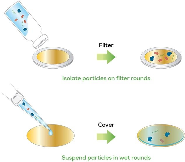
Particles can be isolated from the drug product by membrane filtration. Gold-coated filter rounds maximize contrast between the particles and background, so no particles are missed. Amorphous particles can be analyzed in suspension. Thanks to the flexibility of microscopy, imaging and counting particles in a liquid suspension is no problem. Powders and creams can be spread in a monolayer for analysis.
Make it count
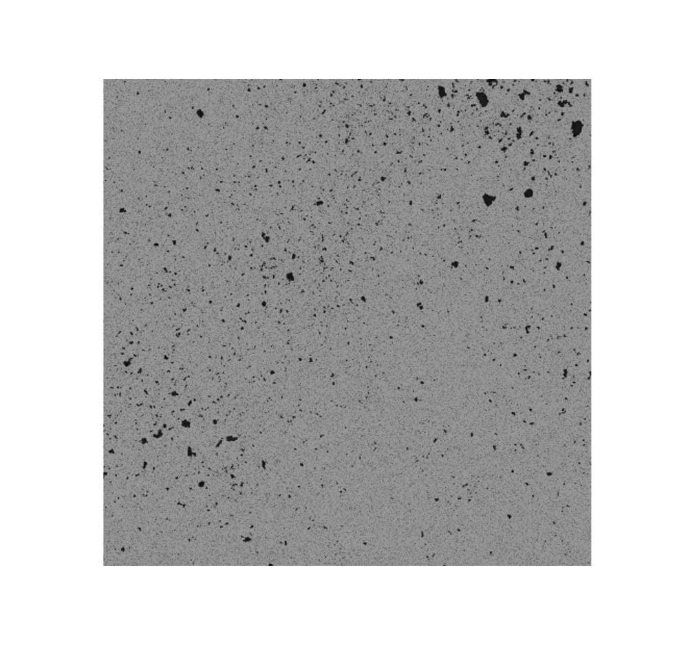
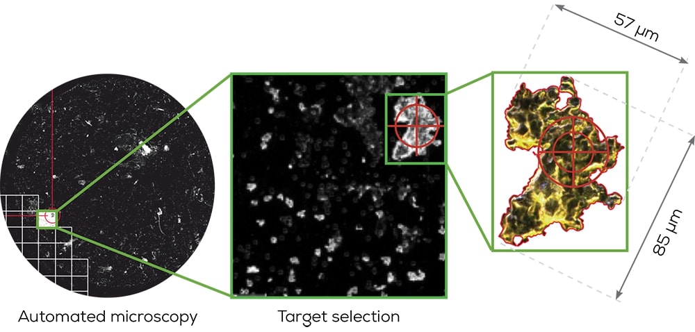
A series of images are collected and stitched together, creating a mosaic image spanning a large area. A predefined threshold can be used to differentiate particles versus background, resulting in a particle count for the sample.
Size them up
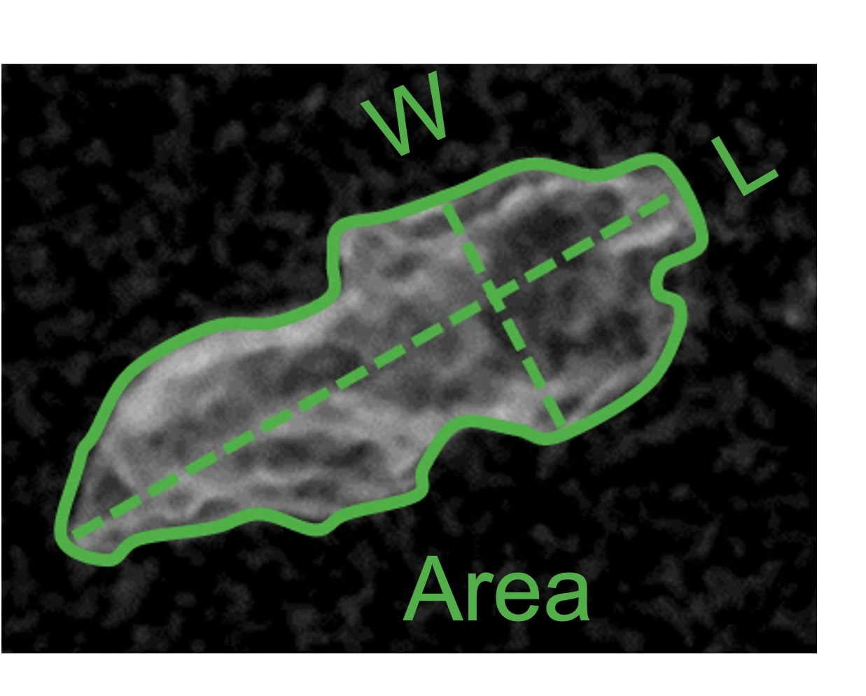
With images collected of each particle, the size and shape can be fully characterized. Automated microscopy measures each dimension of a particle and calculates various morphological parameters, such as elongation, fibrosity, or equivalent circle diameter.
Get organized
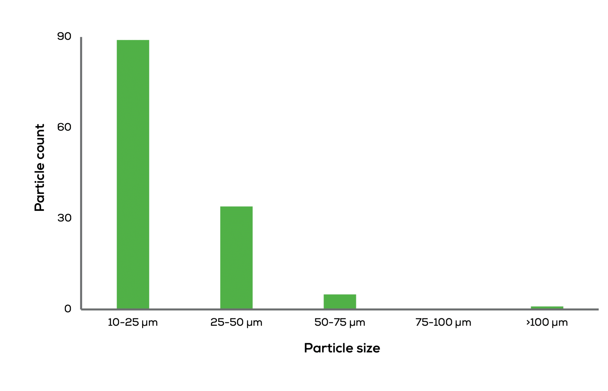
Particles can be binned based on size or other morphological parameters, so you can go back and focus on the particles that matter the most.
Hound
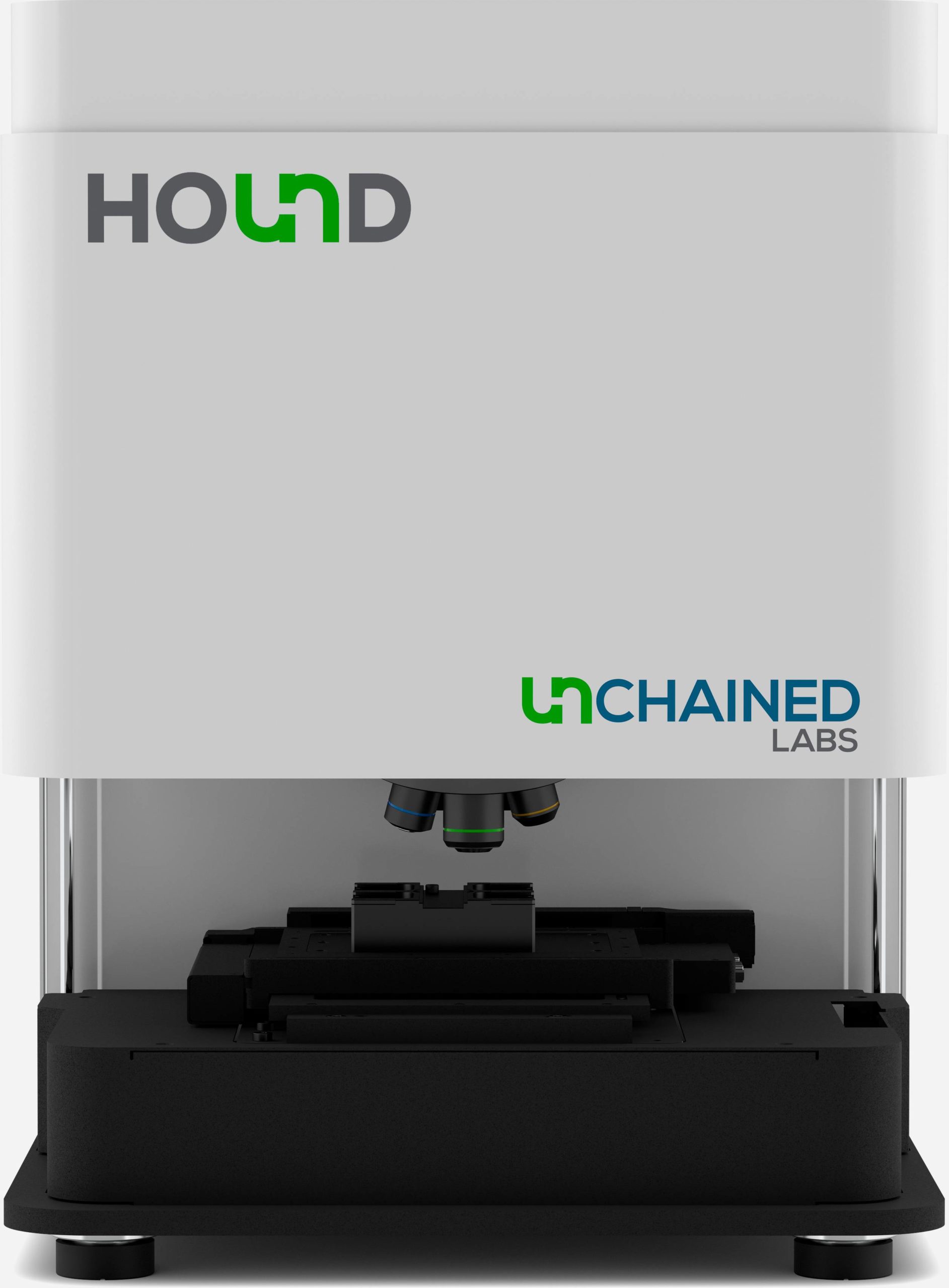
Combining automated microscopy, Raman spectroscopy, and Laser Induced Breakdown Spectroscopy (LIBS) in a single instrument, Hound enables manual and automated identification of visible and subvisible particles across a wide range of chemical compositions.
Ready for more?
Biopharma scientists can now use the right tool for microscopy particle size analysis and identification challenges with an instrument that combines laser induced breakdown spectroscopy, Raman spectroscopy, and light microscopy. Have a question or ready to find out more? Here’s the best way to reach us:


